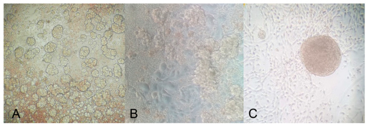
Figure 6.
A few hours after cell isolation, a strong component of spheroids was visible in culture (A); after 24 h, although the presence of spheroids in suspension was still massive, adherent cells became predominant (B); after the first passage in culture, cells showed the capacity to agglomerate and return in suspension as spheroids, particularly when the cells were seeded onto the HA matrix (C). The mounted coverslips were examined under a Leica AF6500 microscope using the LAS software (Leica). Original magnification: 200×.

