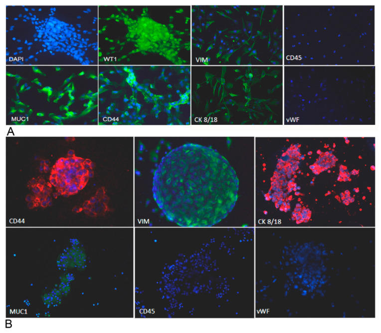
Figure 7.
Ovarian cancer adherent cells and spheroids were characterized for the expression of different markers. We evaluated the expression of several markers for ovarian cancer cells by immunofluorescence, upon fixation and permeabilization. Adherent cells (A) appeared positively stained for vimentin, cytokeratin 8/18, mucin 1, CD44 and WT-1 and negatively stained for von Willebrand factor and CD45, excluding endothelial cell and blood cell contamination. Spheroids (B), after cytocentrifugation and fixation, were stained for vimentin, cytokeratin 8/18, mucin 1, CD44 and WT-1. Nuclei were stained with DAPI. Original magnification: 100×.

