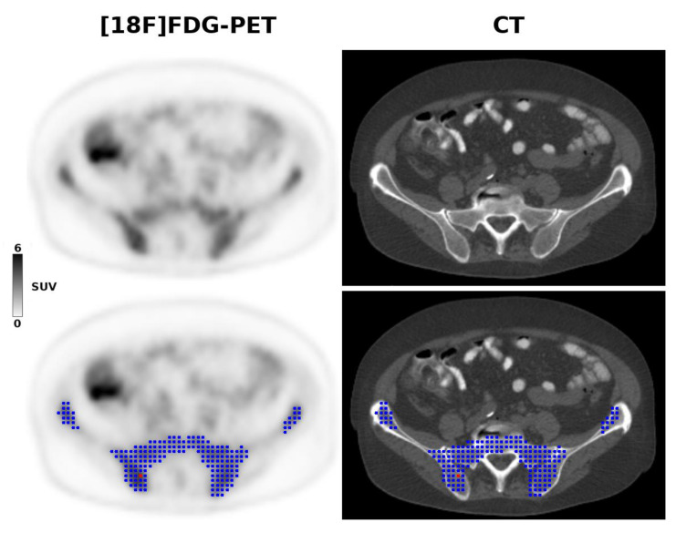
Figure 2.
A 68-year-old patient with stage IV mantle cell lymphoma due to biopsy-proven bone marrow involvement. The [18F]FDG-PET 3D radiomic analysis is based on the metabolic tumor volume (blue) within the pelvis, constructed using the previously recommended 41% SUVmax threshold, and controlled by CT anatomy. The red dot shows the voxel with the highest SUV (i.e., the SUVmax).

