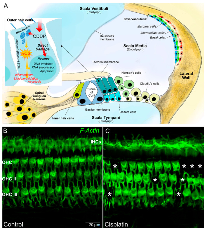
Figure 1.
In (A) a schematic representation of a transverse section of the basal part of a mammalian cochlea. Organ of Corti cellular organization: one inner hair cell (IHC) and three outer hair cells (OHCs) are represented on either side of the tunnel of Corti. The tectorial membrane, floating in endolymph, caps the tallest stereocilia of the hair cells. The IHC is surrounded by supporting cells, whereas OHCs are anchored to the Deiters’ cells, their lateral membrane in direct contact with a fluid called endolymph, which fills the tunnel of Corti. The lateral wall is constituted by the stria vascularis and spiral ligament. In (B), representative images are shown of surface preparations of the organ of Corti stained for F actin, used to visualize the stereociliary arrays and cuticular plates of hair cells. The dark spots indicated by asterisks in (C) show OHC loss after a single dose of cisplatin in the rat model.

