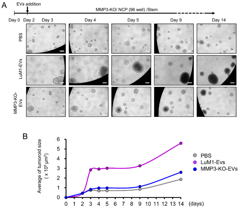
Figure 5.
The addition of MMP3-rich EVs accelerated the in vitro tumorigenesis of MMP3-KO cells. MMP3-KO tumoroids were treated with PBS, LuM1-EVs, or MMP3-KO-EVs at a final concentration of 5 μg/mL in the NCP-based 3D culture with the stemness-enhancing medium. (A) Experimental scheme (top) and representative photomicrographs (bottom) of tumoroid maturation at the indicated timepoints. Scale bar, 100 µm. (B) A time plot graph showing the average size of the MMP3-KO tumoroids following the different treatments over the indicated time points. See the next figure for statistical analysis.

