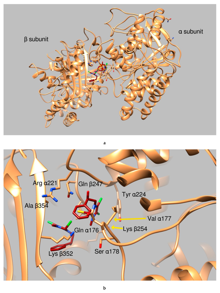
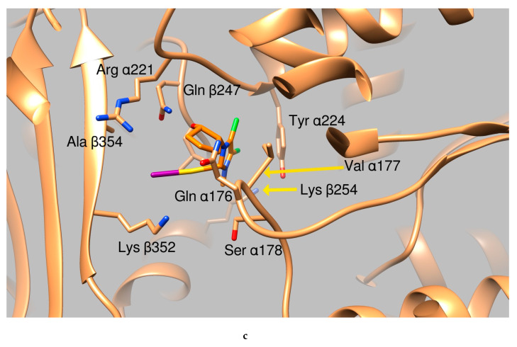
Figure 3.
Graphical representation of AuL4 and AuL7 binding to the tubulin α:β interface. (a) Ribbon representation of the protein dimeric interface (tanned ribbons), bound ligands are reported as sticks. Residues involved in the binding are properly labeled. (b) Tubulin dimeric interface in complex with AuL7 (protein is represented in tanned ribbons, amino acids involved in ligand binding are properly labeled, AuL7 is drawn as purple sticks). (c) Close up of AuL4 (orange sticks) binding.


