Figure 5.
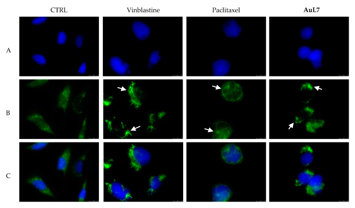
Immunofluorescence studies. MDA-MB-231 cells were treated with AuL7, Vinblastine, Paclitaxel (used at their IC50 values) or with a vehicle (CTRL) for 24 h. After treatment, the cells were methanol fixed, incubated with primary and secondary antibodies, stained with 4′,6-diamidino-2-phenylindole (DAPI) and observed and imaged under the inverted fluorescence microscope at 63× magnification (see Materials and methods). Vehicle-treated cells (CTRL) exhibited a normal arrangement and organization of the cytoskeleton. Microtubules of MDA-MB-231 cells treated with vinblastine and paclitaxel showed an irregular arrangement and organization: particularly, tubulin crystal formation (white arrows, panel B, vinblastine) and tubulin bundles and thicken fibers (white arrows, panel B, paclitaxel) can be noted for vinblastine and paclitaxel respectively. Treatment with AuL7 shows a vinblastine-like mode of action (white arrows, panels B, AuL7). Panels A: DAPI, excitation/emission wavelength 350 nm/460 nm; panels B: β-tubulin (Alexa Fluor® 488) excitation/emission wavelength 490 nm/515 nm; panels C show a merge. Representative fields are shown.

