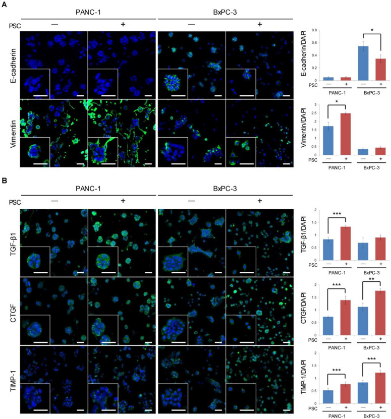
Figure 2.
Expression of epithelial-mesenchymal transition (EMT)-related proteins in tumor spheroids (TSs) under PSC co-culture conditions. (A) Expression of EMT marker proteins E-cadherin and vimentin. (B) Expression of EMT-inducing factors TGF-β1, CTGF, and TIMP-1. Protein expression levels were normalized by nuclear staining with DAPI. Optical sections were acquired at 6 µm (10×) or 2 µm (40×) intervals and stacked into a z-projection. Cells were grown for 5 days in collagen-supported microchannel chips. Three fields covering 80% of the effective area in each channel were imaged per experiment and subjected to analysis. Data were obtained from three separate independent experiments. Scale bar: 50 µm (A, B). * p < 0.05, ** p < 0.01, *** p < 0.005.

