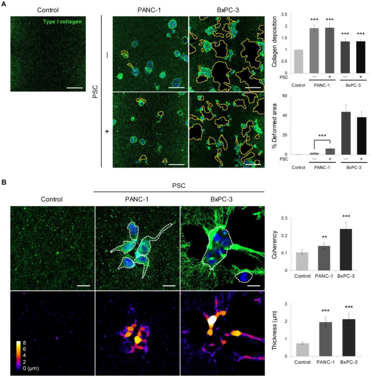
Figure 4.
Differential remodeling of the collagen matrix between PANC-1 and BxPC-3 cells. (A) Deposition and spatial distribution of PANC-1 and BxPC-3 cells with or without PSC co-culture. Scale bar: 100 µm. (B) Changes in collagen structure and organization with respect to coherence and thickness. Scale bar: 20 µm. Control: cell-free matrix. Three fields were imaged per experiments and more than 10 regions of interest were selected for the experiment and subjected to analysis. Data were obtained from three separate independent experiments. ** p < 0.01, *** p < 0.005.

