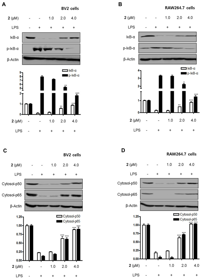
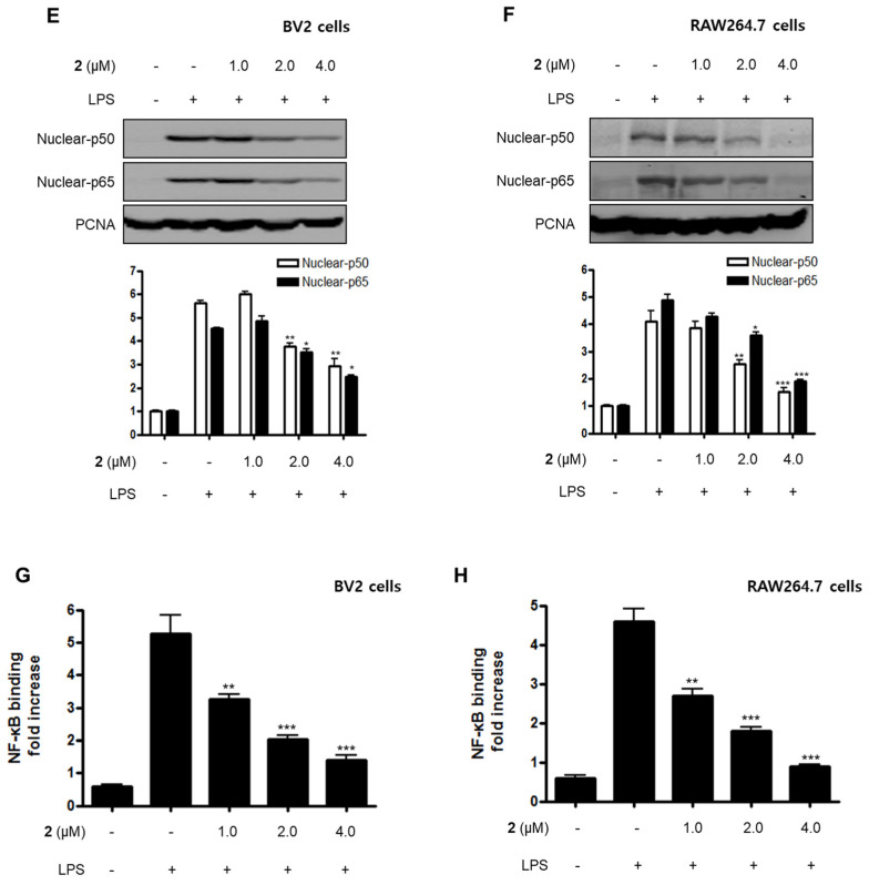
Figure 4.
Effect of compound 2 on the activation of the NF-κB pathway in LPS-induced BV2 and RAW264.7 cells. After pre-treatment with compound 2 (1.0, 2.0, and 4.0 μM) for 3 h, the cells were stimulated with LPS for 1 h. (A–F) Proteins were obtained, and specific anti-IκB-α, anti-p- IκB-α, anti-p65, and anti-p50 antibodies were employed for western blot analysis. Representative blots of three independent experiments are shown. Band intensity was quantified by densitometry and normalized to β-actin for the cytoplasmic fraction and to proliferating cell nuclear antigen (PCNA) for the nuclear fraction. (G, H) NF-κB binding activity in the nuclear fraction was determined using an NF-κB ELISA kit by Active Motif (* p < 0.05; ** p < 0.01; *** p < 0.001) and then compared to that of the LPS-treated group.


