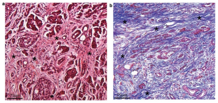
Figure 1.
(a) Hematoxylin and Eosin (H&E stain) of a surgically resected pancreatic ductal adenocarcinoma showing small and medium glands with irregular morphology embedded in dense, desmoplastic stroma (highlighted with black asterisks). (b) Trichrome stain of surgically resected pancreatic ductal adenocarcinoma highlighting severe desmoplasia and dense matrix that appears as linearized ribbons of blue stain (collagen fibers) (highlighted with black asterisks).

