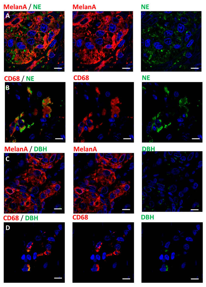
Figure 2.
Norepinephrine expression in melanoma tumors: (A) Photographs showing melanoma cells stained for MelanA (red) antigen expressing norepinephrine (NE, green). (B) Photograph showing CD68 positive macrophages (red) carrying norepinephrine granules (NE, Green). (C) Photographs showing the expression of dopamine beta-hydroxylase (DBH, green) by melanoma cells (MelanA, red). (D) Expression of DBH (green) in a population of macrophages (CD68, red). Scale bars 10 µm.

