Figure 6.
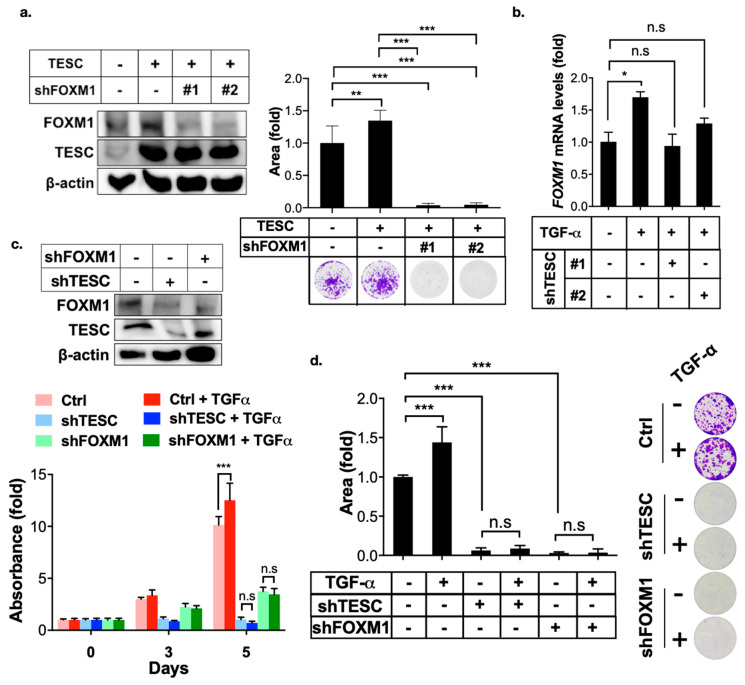
TESC mediates TGF-α-induced FOXM1 expression. (a) Clonogenic assays were employed to assess the effect of FOXM1 silenced on TESC-overexpressed HUCCT1 cells. HUCCT1 cells were first infected with lentiviral vectors encoding TESC or vector control for 5 days, followed by infection with lentiviral vectors encoding shTESC or scrambled control for 2 weeks. The levels of TESC and FOXM1 were determined by immunoblotting (left). Colonies were stained with crystal violet and quantified (upper right). Representative plates were photographed (bottom right). ** p < 0.01, *** p < 0.001. (b) qPCR analysis of FOXM1 expression in HUCCT1 cells infected with or without shTESC in the presence TGF-α (50 ng/mL) for 4 h. * p < 0.05, n.s.: no significance. (c) MTT assay of proliferation of HUCCT1 cells infected with shTESC as well as shFOXM1 or control vector in the presence or absence of TGF-α (50 ng/mL). The levels of TESC and FOXM1 were determined by immunoblotting (upper). *** p < 0.001, n.s.: no significance. (d) Clonogenic assays of HUCCT1 cells infected with lentiviral vectors encoding shTESC, shFXOM1, or scrambled control in the presence or absence of TGF-α (50 ng/mL) for 2 weeds. Left: Colonies were stained with crystal violet and quantified. Right: Representative plates. *** p < 0.001, n.s.: no significance.

