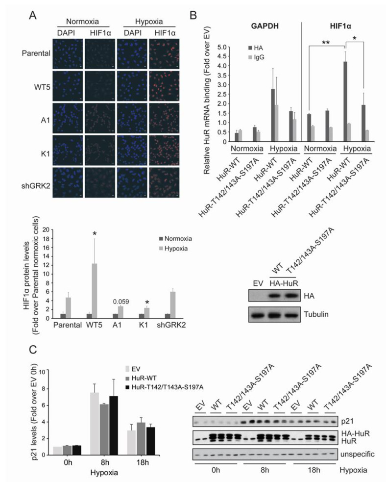
Figure 4.
GRK2 kinase activity is required for HuR-induced upregulation of HIF1α. (A) HeLa stable cell lines were cultured under hypoxia for 2 h and HIF1α levels were analyzed by immunofluorescence. Values are mean ± SEM (fold-change over parental cells in normoxia) from 3–4 independent experiments. * p < 0.05 (Student’s t-test) comparing the changes in hypoxic HIF1α levels in Hela stable cells lines, with those observed in hypoxic parental cells. Representative images are shown. Scale Bar = 10 µm. (B) HeLa cells were transiently transfected with pcDNA3 as empty vector (EV), HA-HuR WT, or HA-HuR-T142/143A-S197A, and cultured for 4 h under hypoxia. Total lysates were then immunoprecipitated with either an HA antibody or control IgGs. RNA was purified from immunoprecipitates and used for qRT-PCRs. GAPDH was used as an endogenous control. The graph shows fold differences in transcript abundance (mean ± SEM in two independent experiments performed in triplicates). ** p < 0.01 hypoxic vs. normoxic WT; * p < 0.05 hypoxic triple mutant vs. hypoxic WT (two-way ANOVA). The expression levels of the different HA-HuR proteins analyzed by immunoblotting are shown below. (C) HeLa cells were transiently transfected with pcDNA3 as empty vector (EV), HA-HuR WT, or HA-HuR-T142/143A-S197A, and was cultured under hypoxia for the indicated times. p21 and endogenous and overexpressed HuR levels were analyzed by immunoblot. A non-specific band was used as the loading control. Values are mean ± SEM from two independent experiments. Detailed information about the Western blots can be found in Figure S5.

