Figure 5.
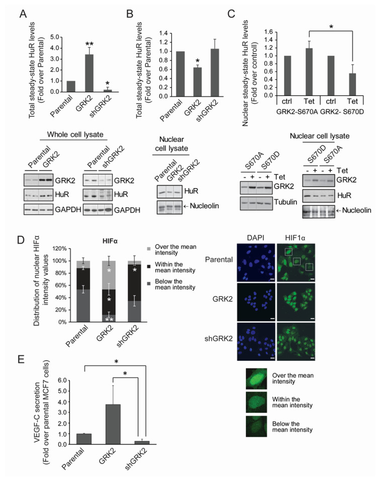
GRK2 modulates HuR levels and cytoplasmic shuttling, HIF1-α distribution, and VEGF-C secretion in luminal breast cancer cells. (A,B) Whole lysates or nuclear extracts from MCF7 cells stably expressing GRK2-wt or shGRK2 were used to analyze the HuR levels by immunoblot. Values are mean ± SEM from 3–4 independent experiments. * p < 0.05 ** p < 0.01 (Student’s t-test). Representative blots with loading controls are shown. (C) MCF7 stable inducible cell lines were treated with tetracyclin (Tet) to induce the expression of the indicated GRK2 mutants. HuR nuclear levels were analyzed by immunoblot. Values are mean ± SEM from two independent experiments. * p < 0.05 (Student’s t-test). Representative blots with loading controls are shown. (D,E) HIF1-α distribution and VEGF-C secretion levels were analyzed in parental MCF7 cells or in derived cells lines stably overexpressing GRK2 or a silencing construct (shGRK2) of the kinase. (D) HIF1-α and DAPI staining was analyzed by immunofluorescence; distribution of intensity values is shown. Values are distribution percentages of cells with HIF1-α intensity values over, within, or below the mean intensity. Representative images are shown. * p < 0.05; ** p < 0.01 (Student’s t-test) when each category (over, within, below) in MCF7-GRK2 and MCF7-shGRK2 cells is compared to parental ones. Scale Bar = 25 µm. (E) ELISA-measured VEGF-C secretion values are shown (mean ± SEM from 3 independent experiments). * p < 0.05 (Student’s t-test). Detailed information about the Western blots can be found in Figure S7.

