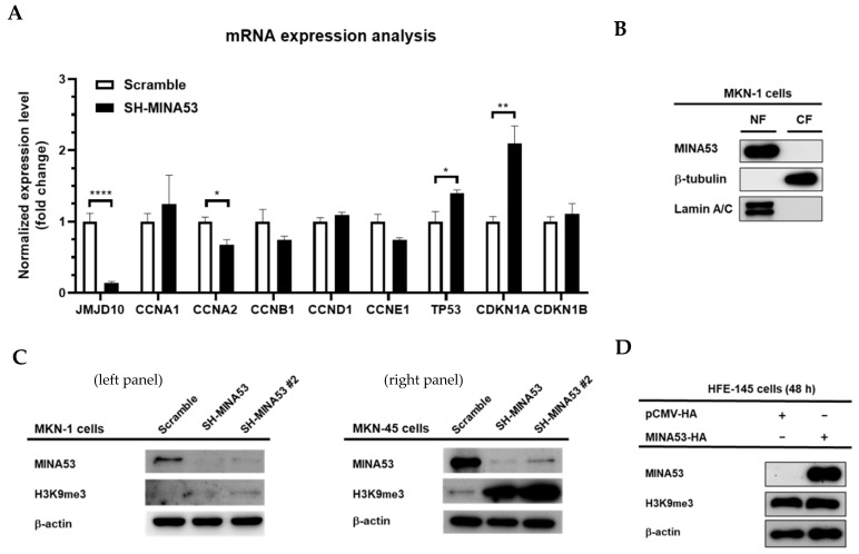
Figure 5.
Molecular signature of MINA53 in gastric cancer cell lines. (A) mRNA expression analysis of JMJD10 in MINA53-silenced MKN-1 cells. (B) MINA53 mainly localized in the nuclear fraction; Lamin A/C and β-tubulin were used as loading controls for nuclear and cytosolic fractions, respectively. (C) Analysis of H3K9me3 level in MINA53-silenced MKN-1 cells (left panel) and MKN-45 cells (right panel). (D) Overexpression of MINA53 in human gastric normal cell lines (HFE-145) does not affect the H3K9me3 level. The whole Western blot images please find in Figure S1.

