Figure 1.
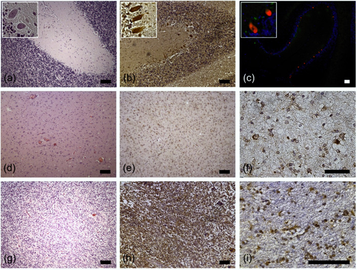
Representative examples of RPS27 immunohistochemistry. (a) H&E, (b) RPS27-DAB, and (c) RPS27 fluorescence staining of normal cerebellar tissue. Purkinje cells were positive for RPS27 staining (red). DAPI = blue. Insets’ objective magnification 40×. (d) H&E, (e) and (f) RPS27-DAB staining of an exemplary WHO grade II glioma. (g) H&E, (h) and (i) RPS27-DAB staining of an exemplary glioblastoma. Scale bars = 100 µm.

