Figure 2.
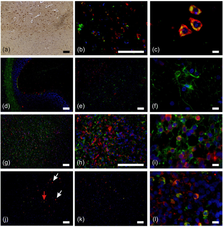
Fluorescence double staining of normal brain and astrocytic tumor specimens. (a) RPS27-DAB staining of normal brain tissue. Positive cells were concentrated in the grey matter of the frontal cerebrum. (b) and (c) neurons (NeuN = green, RPS27 = red) in the frontal cerebrum. (d) Overview of the cerebellum double stained for RPS27 (red) and astrocytes (glial fibrillary acidic protein, GFAP = green). (e) Neurons and astrocytes in the frontal cerebrum (RPS27 = red, GFAP = green) and (f) astrocytes in 100× magnification (RPS27 = red, GFAP = green). (g–i) Glioblastoma (GBM) cells in the cerebellum. RPS27 = red, GFAP = green. (j) Diffuse IDH1 R132H mutant GBM. (RPS27 = red, IDH1 R132H = green). Red arrow = RPS27/IDH R132H double positive tumor cell, white arrows = RPS27-positive but IDH1 R132H-negative cells. (k) and (l) GBM double staining for RPS27 (red) and macrophages (CD68 = green). DAPI = blue, scale bars in (c), (f), (i), and (l) = 10 µm, otherwise scale bars = 100 µm.

