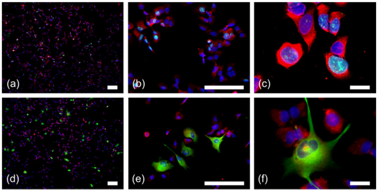
Figure 4.
RPS27 staining in conjunction with Ki67 and GFAP positivity in GBM stem-like cells. (a–c) Immunofluorescence double staining for RPS27 (red) and Ki67 (green). (d–f) Immunofluorescence double staining for RPS27 (red) and GFAP (green). DAPI = blue, scale bars in (c) and (f) = 20 µm, otherwise scale bars = 100 µm. For the single color channels see Figure S2.

