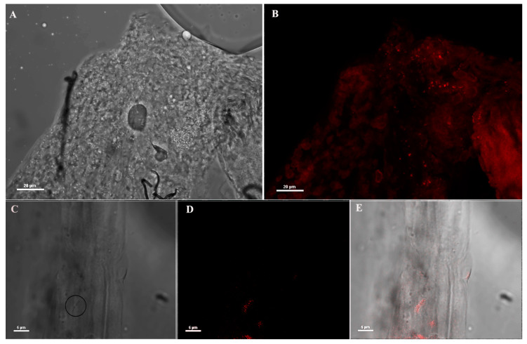
Figure 1.
Immunofluorescence assay (IFA) on mosquito midguts using anti-Asaia monoclonal antibody (mAb). (A,B) Bright field microscopic and immunofluorescence images (40X) of male midgut using anti-Asaia mAbs; (C) different magnification (100X) of the tissues highlighting bacteria cells with the circle; (D) the corresponding immunofluorescent staining showing red-labelled bacteria recognized by the mAb; and (E) the superimposed image for localization. Scale bar = 20 μm (A,B) and = 5 μm (C–E).

