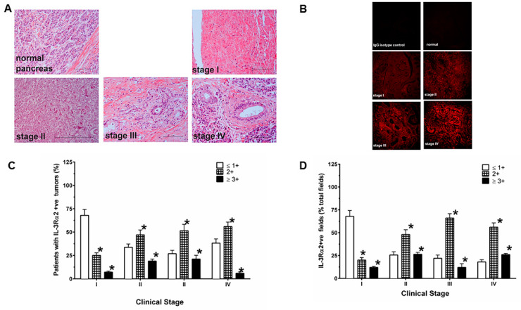
Figure 2.
IL-13Rα2 expressing tumor cells in different clinical stages. (A) H&E staining of PDAC samples with stage I-IV tumors (B) IHC (immunohisto-chemistry) of IL-13Rα2 expression in PDAC with different stages and normal pancreas. (C) The extent of IHC staining in PDAC was evaluated at four levels between 0 and 3+ according to the intensity of immunostaining. (D) Percent positive fields expressing IL-13Rα2 were counted in samples with different grades. Normal pancreas showed negative staining for IL-13R2e expression. The samples were viewed at 200× magnification. * p = 0.0001.

