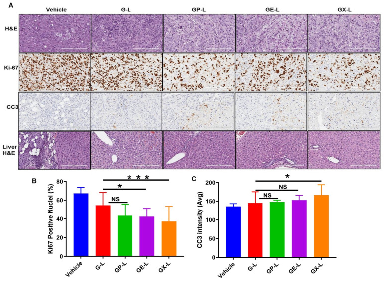
Figure 7.
H&E, Ki67, and CC3 staining of tumor sections and liver H&E obtained from respective treatment groups. (A) Representative images of the H&E- and Ki67-stained tumor sections from different treatment groups displayed the comparatively higher antiproliferative activity of dual drug-loaded liposomes than gemcitabine-loaded liposomes and untreated. Quantification of Ki67-positive nuclei (B) and average CC3 positive intensity (avg) (C) of tumor sections. * and *** denotes p < 0.05 and p < 0.001 compared to G-L, respectively.

