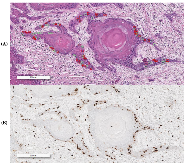
Figure 2.
An overlapping field with SCC (squamous cell carcinoma) obtained from the Ki67 immunostained H&E decolored sample. (A) H&E (hematoxylin and eosin) (squares indicate some of the selected cells for feature analysis); (B) Ki67/MIB1 IHC (immunohistochemical). Red and green squares, respectively, represent positive and negatively annotated cells.

