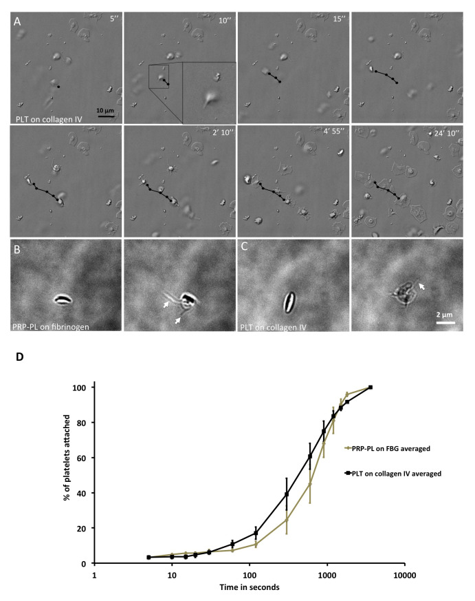
Figure 1. Observations of filopodia on platelets in solution on fibrinogen and collagen IV.
( A) DIC, time-lapse microscopy of platelets attaching to collagen IV at different time points. Black arrow denotes the platelet in question. Scale bar: 10 μm. ( B) Phase-contrast microscopy of PRP-PL on fibrinogen. Left: Discoid shape; Right: Platelet with filopodial extension. Scale bar: 5 μm. ( C) Phase-contrast microscopy of PLT on collagen IV. Left: Discoid shape; Right: Platelet with filopodial extension. Scale bar: 5 μm. ( D) Platelet attachment to collagen IV or fibrinogen in percentage (%) over time. Time in seconds, on a logarithmic scale. Platelets on collagen IV attach faster that on fibrinogen. Wilcoxon rank-sum test, p-value < 0.015. N platelets: FBG=121, collagen IV=274. Error bars show Standard Error.

