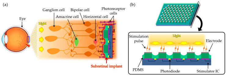
Figure 1.
Schematic illustrations of microelectrodes for subretinal stimulation. (a) The subretinal implant is placed between the outer nuclear layer (ONL) and the retinal pigment epithelium (RPE) to stimulate non-spiking neurons. (b) Stimulation pulses are generated when photodiodes in the implant convert light into electrical signals.

