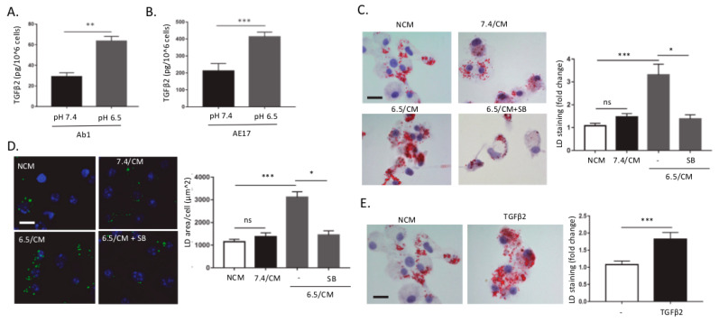
Figure 1.
TGF-β2-dependent lipid droplet accumulation in dendritic cells in response to the acidic mesothelioma milieu. (A,B) Control (pH 7.4) and acidosis (pH 6.5)-adapted Ab1 (A) and AE17 (B) mesothelioma cells were grown for 48 h, and active TGF-β2 secretion was assayed using ELISA. (C–E) Dendritic cells (DCs) were incubated with non-conditioned medium (NCM) or treated for two days either with conditioned medium (CM) from mesothelioma cells maintained at pH 7.4 or pH 6.5 (7.4/CM and 6.5/CM, respectively) (C,D) or with 4 ng/mL recombinant TGF-β2 (E). In some experiments, DCs were also exposed to 5 µM SB-431542. Representative pictures of lipid droplet (LD) content as determined using Oil Red O (ORO) (scale = 20 µm) (C,E) or BODIPY 495/503 staining (scale: 20 µm, green: BODIPY 495/503, blue: DAPI) (D) are shown together with quantification of the cellular area covered by LDs (n = 3, * p < 0.05, ** p < 0.01, *** p < 0.001; ns = non-significant).

