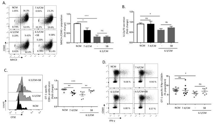
Figure 4.
CD8+ T cell response is altered upon dendritic cells (DCs) exposure to the mesothelioma acidic milieu. DCs were incubated with non-conditioned medium (NCM) or treated for 2 days either with conditioned medium from AE17 or Ab1 mesothelioma cells maintained at pH 7.4 or pH 6.5 (7.4/CM and 6.5/CM, respectively). (A,B) Effects of 6.5/CM with or without 5 µM SB-431542 on MHCII+/CD40+ surface expression as determined by flow cytometry (A) and pro-inflammatory interleukin-12 (IL-12p70) secretion as detected by ELISA (B). (C,D) Effects of 6.5/CM with or without 5 µM SB-431542 on OT-1 specific CD8+ proliferation, as measured using carboxyfluorescein succinimidyl ester (CFSE) dilution (C) and activation, as evaluated by determining CD69+/IFN-γ+ frequency (D). The charts and histogram presented in this figure are representative of at least three different experiments. * p < 0.05, ** p < 0.01, *** p < 0.001; ns = non-significant.

