Fig. 2.
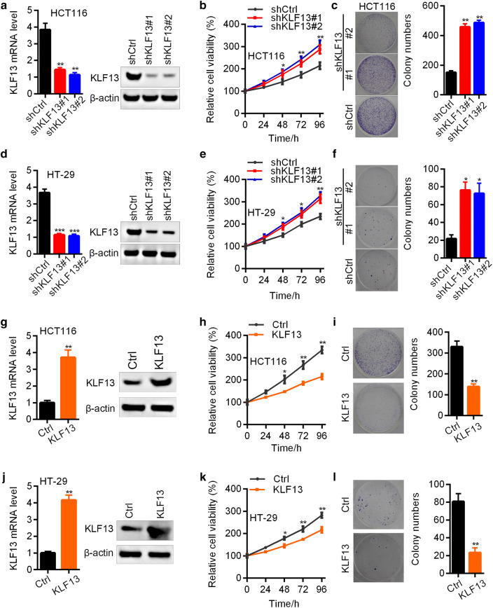
KLF13 suppresses the proliferation of CRC cells. a–c shCtrl, shKLF13#1 and shKLF13#2 HCT116 cells were subjected to RT-qPCR and Western blot analysis of KLF13 (a), CCK8 analysis of cell proliferation (b), and colony formation assay (c Left, representative images; Right, quantification results). *p < 0.05, **p < 0.01. d–f shCtrl, shKLF13#1 and shKLF13#2 HT-29 cells were subjected to RT-qPCR and Western blot analysis of KLF13 (d), CCK8 analysis of cell proliferation (e), and colony formation assay (f Left, representative images; Right, quantification results). *p < 0.05, **p < 0.01, ***p < 0.001. g–i Ctrl and KLF13 overexpressed HCT116 cells were subjected to RT-qPCR and Western blot analysis of KLF13 (g), CCK8 analysis of cell proliferation (h), and colony formation assay (i Left, representative images; Right, quantification results). *p < 0.05, **p < 0.01. j–l Ctrl and KLF13 overexpressed HT-29 cells were subjected to RT-qPCR and Western blot analysis of KLF13 (j), and CCK8 analysis of cell proliferation (k), and colony formation assay (l Left, representative images; Right, quantification results). *p < 0.05, **p < 0.01

