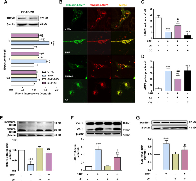
Fig. 7.
TRPM2 channel activation mediates SiNPs-induced lysosome impairment and blockage of autophagic flux in BEAS-2B cells. a Western blotting analysis of TRPM2 expression in cells (top) and single cell imaging analysis using Fluo-3 of intracellular free Ca2+ levels in cells treated with SiNPs (100 μg/mL) for 0.5, 1, 2, 3 h in the absence or presence of PJ34 (10 μM) and compound A1 (10 μM) (bottom). b Representative confocal microscopic images showing mApple-LAMP1-pHluorin fluorescence in cells under control condition or after treated with SiNPs (100 μg/mL) for 24 h in the absence or presence of compound A1 (10 μM). Scale bar = 10 μm. c-d Mean number of puncta in each cell under indicated onditions shown in B, from 30 cells for each condition. e-g Western blotting analysis the levels of CTSD (e), LC3 (f) and SQSTM1 (g) in cells under control condition or after treatment with SiNPs (100 μg/mL) in the absence or presence of compound A1 (10 μM), from three independent experiments. *P < 0.05, **P < 0.01, ***P < 0.001 compared to the control cells, and #P < 0.05, ##P < 0.01 compared to cells treated with SiNPs alone

