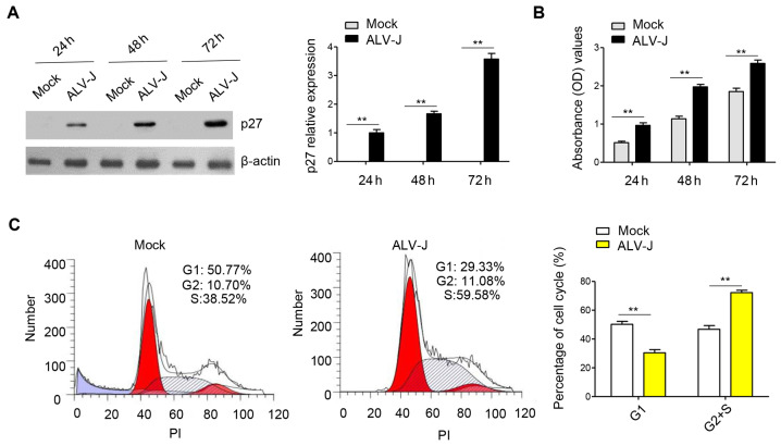
Figure 1.
ALV-J infection in DF-1 cells promotes proliferation and the cell cycle. (A) DF-1 cells were infected with ALV-J GD1109 strain with a multiplicity of injection of 1. Cells were harvested at 24, 48 and 72 h for western blot analysis with antibodies against p27 and β-actin. p27, the capsid protein of ALV-J, was used to measure the viral replication in DF-1 cells. Densitometry analysis was conducted to compare the viral replication in ALV-J-infected group compared with the mock group. (B) Cell proliferation analysis was performed using the Cell Counting Kit-8 assay in DF-1 cells infected with ALV-J, which were examined at 24, 48 and 72 h after transfection. The mock group is the control group. (C) Cell cycle assay was performed in DF-1 cells infected with ALV-J for 48 h, stained with PI and evaluated with a FACSCalibur flow cytometer compared with the mock group. Data are presented as the mean ± SD of three independent experiments. **P<0.01. ALV-J, avian leukosis virus; PI, propidium iodide; OD, optical density.

