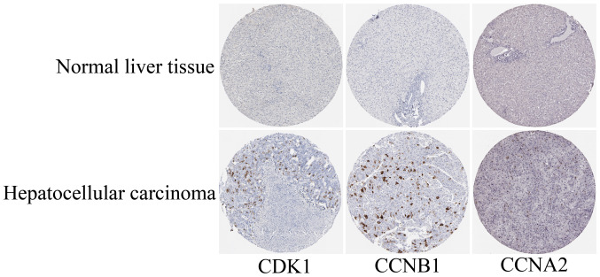
Figure 6.
Representative histological images from the Human Protein Atlas database (https://www.proteinatlas.org/). The normal liver tissue with staining for CDK1 was from a female subject aged 29 years (patient ID: 1899; staining: Not detected; intensity: Negative; quantity: Negative; location: None; magnification, not available) and the HCC tissue was from a female patient aged 52 years (patient ID: 2399; staining: Medium; intensity: Strong; quantity: <25%; location: Cytoplasmic/membranous nuclear; magnification, not available). The normal liver tissue with staining for CCNB1 was from a female subject aged 32 years (patient ID: 1846; staining: Not detected; intensity: Negative; quantity: Negative; location: None; magnification, not available) and the HCC tissue was from a male patient aged 49 years (patient ID: 929; staining: Medium; intensity: Strong; quantity: <25%; location: Cytoplasmic/membranous; magnification, not available). The normal liver tissue with staining for CCNA2 was from a female subject aged 50 years (patient ID: 2251; staining: Not detected; intensity: Negative; quantity: Negative; location: None; magnification, not available) and the HCC tissue was from a female patient aged 73 years (patient ID: 5192; staining: Low; intensity: Moderate; quantity: <25%; location: Nuclear; magnification, not available). CDK1, cyclin-dependent kinase 1; CCNA2, cyclin A2; HCC, hepatocellular carcinoma.

