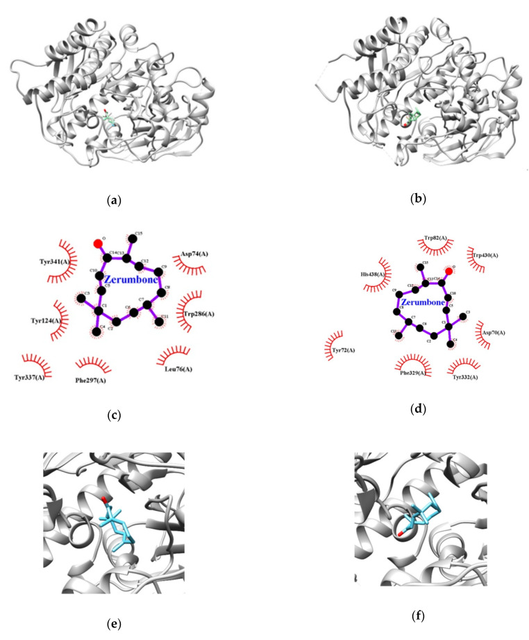
Figure 3.
Representations of the best poses of zerumbone in docking with AChE (a) and BChE (b). Zoomed in view of the zerumbone binding site at AChE (c) and BChE (d). The hydrophobic interaction between AChE (e), BChE (f), and zerumbone. The hydrophobic interactions are depicted as red dashed semicircles.

