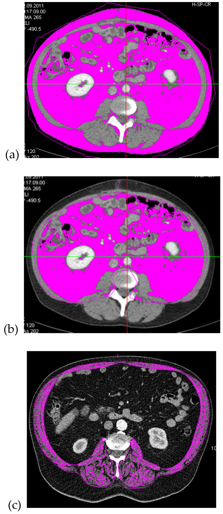
Figure 1.
Example of a computed tomography (CT)-scan with the area-based, densitometric quantification of adipose tissue (threshold: −190 to −30 HU) measured at spinal level L3/4: regions of interest (ROI) containing total fat area (TFA) (a) and visceral fat area (VFA) (b); and an example of the densitometric quantification of muscle area, also measured at spinal level L 3/4 with an ROI containing the muscle tissue of the abdominal, dorsal and psoas muscles (threshold: 40 to 100 HU) (SMM, (c). Computed tomography (CT); Hounsfield units (HU); regions of interest (ROI); skeletal muscle mass (SMM); total fat area (TFA), visceral fat area (VFA).

