Figure 3.

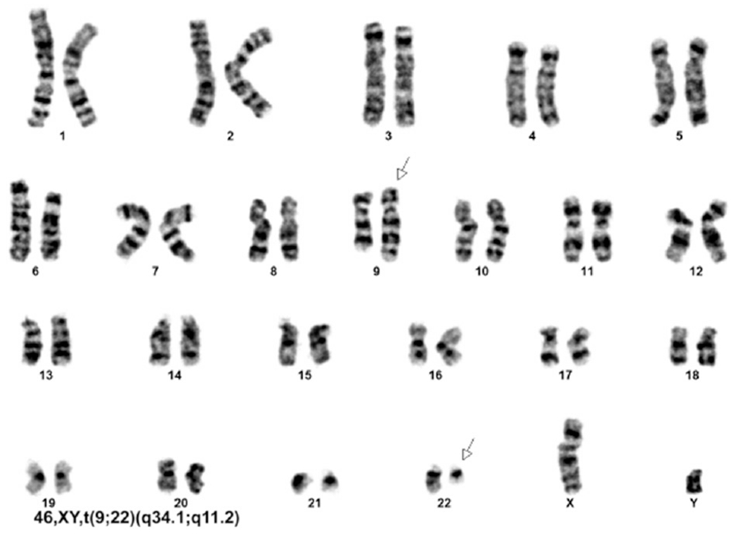
Cytogenetic analysis shows a t(9;22)(q34.1;q11.2) translocation in all 20 cells analyzed. Arrows indicate the abnormal, elongated chromosome 9 (upper arrow) and abnormal, truncated chromosome 22 (lower arrow).

Cytogenetic analysis shows a t(9;22)(q34.1;q11.2) translocation in all 20 cells analyzed. Arrows indicate the abnormal, elongated chromosome 9 (upper arrow) and abnormal, truncated chromosome 22 (lower arrow).