. 2020 May 19;9:e55799. doi: 10.7554/eLife.55799

Figure 5. The FSS-induced ribosome stalling in NR conformation is independent of A-site codon identity.

Histograms show FRET distributions in S6-cy5/L9-cy3 ribosomes programmed with HIV_NS (GAG) (A–B) or HIV_NS (GAG) ∆FSS (C–D) mRNA, respectively. Ribosomes containing P-site N-Ac-Phe-tRNAPhe (A, C) were incubated with EF-Tu•GTP•Tyr-tRNATyr (B, D) for 5 min and imaged after removal of unbound aminoacyl-tRNAs. Yellow lines show individual Gaussian fits of FRET distributions. Black lines indicate the sum of Gaussian fits. N indicates the number of FRET traces compiled into each histogram. The fractions of the ribosome in R and NR conformations are shown above the corresponding 0.4 and 0.6 Gaussian peaks, respectively.

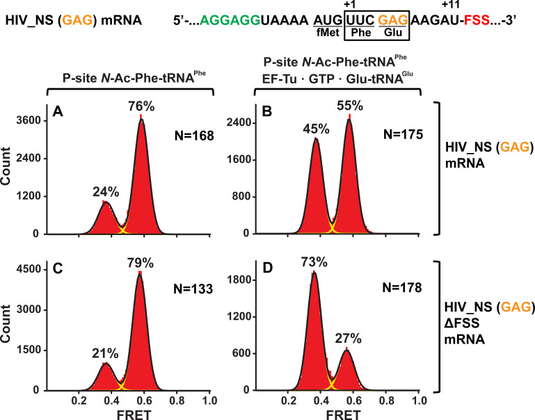
Figure 5.

Figure 5—figure supplement 1. In the absence of slippery sequence, levels of the FSS-induced frameshifting are negligible.

Figure 5—figure supplement 1.

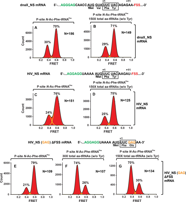
Histograms show FRET distributions in S6-cy5/L9-cy3 ribosomes programmed with the dnaX_NS (A, B), HIV_NS (C, D), or HIV_NS (GAG) ∆FSS (E–G) mRNA, respectively. Ribosomes containing P-site N-Ac-Phe-tRNAPhe (A, C, E) were incubated with 150- (B, D, G) or 30- (F) fold molar excess of total aminoacyl-tRNAs•EF-Tu•GTP (mixture of all aminoacyl-tRNAs except Tyr-tRNATyr) for 5 min and imaged after removal of unbound aminoacyl-tRNA.
Figure 5—figure supplement 2. The FSSs do not stabilize the pretranslocation ribosome in the NR conformation.

Figure 5—figure supplement 2.

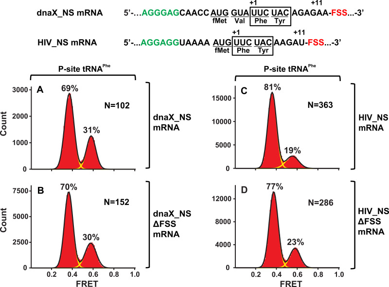
Histograms show FRET distributions in S6-cy5/L9-cy3 ribosomes, which contained deacylated tRNAPhe in the P site and were programmed with the dnaX_NS (A), dnaX_NS ∆FSS (B), HIV_NS (C), or HIV_NS ∆FSS (D) mRNA, respectively.