Fig. 1.

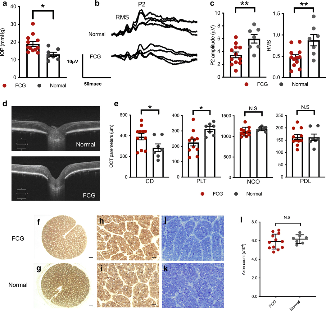
Optic nerve head (ONH) remodeling and functional deficits are evident soon after onset of IOP elevation, prior to axon loss in feline congenital glaucoma (FCG). Mean and SEM are presented throughout; all comparisons between normal (n = 7) and FCG (n = 12) were by unpaired 2-tailed t test. * P < 0.05; ** P < 0.01. a Intraocular pressure (IOP)was significantly higher in FCG than in normal subjects at 10 weeks of age (rebound tonometry; P = 0.01). b, c Peak amplitude of the late positive component (P2) and root mean square (RMS) of the early wavelets of the visual evoked potential (VEP) were significantly lower (P = 0.0058 and 0.0064, respectively) in FCG than controls at 10–12 weeks of age. d Representative ONH spectral domain-optical coherence tomography (SD-OCT) in vivo images of 12-week-old FCG (IOP 15 mmHg, scan quality 10/10) and normal cat (IOP 16 mmHg, scan quality 9/10). e Comparison of SD-OCT-derived quantitative ONH parameters between FCG and normal subjects revealed significantly increased optic cup depth (CD) and reduced pre-laminar tissue thickness (PLT) in FCG (P = 0.044 and 0.011, respectively), whereas width of the neural canal opening (NCO) and posterior displacement of the lamina (PDL) were not significantly different between groups. f–k Axon loss is not an early pathological feature in FCG. Representative optic nerve cross sections from 10- to 12-week-old FCG (f) and normal cats (g). No morphological evidence of axonal damage was observed in FCG (f, h, j) compared to normal control nerves (g, i, k) in either p-phenylenediamine (which stains myelin sheaths) (h, i) or Richardson’s stained sections (j, k). Scale bars = 200 μm (f, g); 20 μm (h–k). l Mean optic nerve axon count in FCG (n = 12) was not significantly different from normal (n = 7; P = 0.447, unpaired 2-tailed t test)
