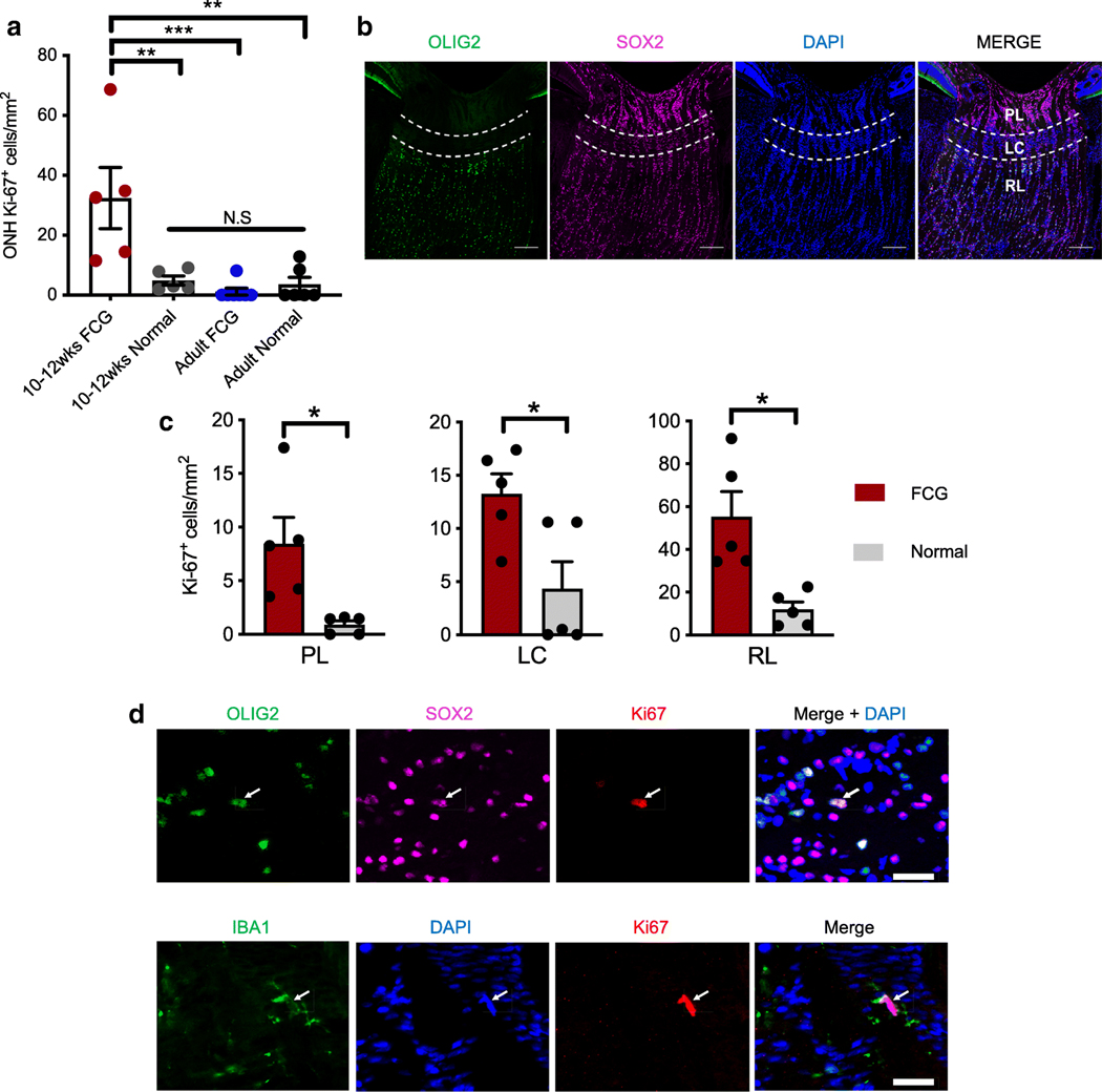
Fig. 3.
Glial cell proliferation in the ONH is a feature of early disease in FCG. a Early glaucomatous ONHs had significantly increased density of proliferating (Ki67-immunopositive) cells compared to ONHs from agematched normal, adult FCG, and adult normal cats (**P < 0.01; ***P < 0.001; ANOVA followed by Tukey’s post-test for multiple comparisons; mean and SEM presented). In contrast, density of proliferating cells in the ONH was not significantly different between groups of adult cats (P = 0.78, n = 6 per group). b Photomicrographs of normal 10–12-week-old feline ONH showing three sub-regions (prelaminar [PL]; lamina cribrosa [LC], and retro-laminar [RL; extending 200 μm posterior to LC]) that are distinguishable based on their morphology and cell populations. OLIG2 oligodendrocyte-lineage marker (green); SOX2 astrocyte and glial progenitor marker (magenta); DAPI nuclear counter staining (blue). Scale bar = 200 μm. c In 10–12week-old cats with FCG, increased density of proliferating (Ki67-positive) cells was observed in each of the three ONH sub-regions, compared to age-matched normal controls (n = 5 per group; *P < 0.05, unpaired t test with Welch’s correction). d In the retrolaminar region of the ONH, Ki67 expression colocalized with SOX2 (magenta; top row) and OLIG2(green;toprow) (top;arrow). Dispersed throughout the ONH, other Ki67-positive cells expressed the microglia/macrophage marker IBA1 (bottom; arrow). Scale bar = 20 μm

