Fig. 5.
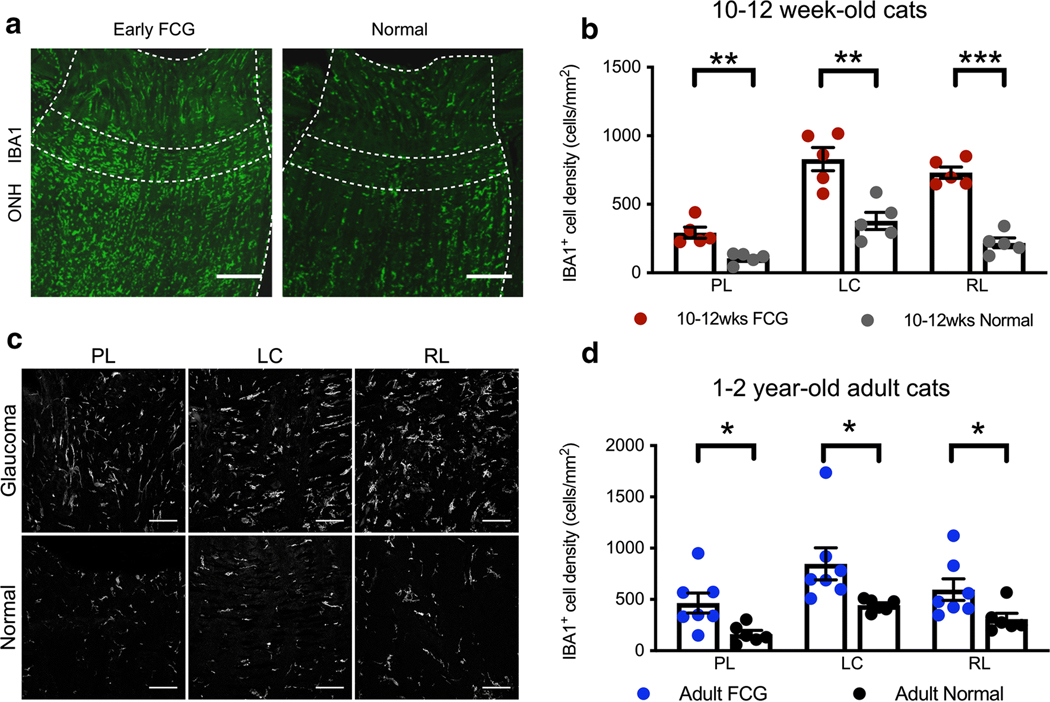
Glaucomatous ONH pathobiology is characterized by microglial/ macrophage activation in both early and chronic FCG. a Photomicrographs illustrate the distribution of IBA1+ microglia in immunolabeled longitudinal sections of early FCG and age-matched control ONHs. Microglia are distributed throughout the ONH in both groups (scale bar = 100 μm). b The density of IBA1+ microglia was significantly increased in all 3 regions of the ONH in early FCG, compared to control subjects (n = 5 per group) (unpaired t test: PL, **P < 0.01; LC, **P < 0.01; RL, ***P < 0.001, respectively). FDR < 0.01 in all three tests. Data presented as mean and SEM. c Representative photomicrographs illustrating IBA1+ cells in prelaminar (PL), lamina cribrosa (LC), and retro-laminar (RL) regions in early glaucomatous and normal age-matched ONH sections. Microgliosis was evident in all sub-regions of the ONH in early FCG (scale bar = 25 μm). d The density of IBA1+ microglia was also increased in all 3 regions of in the ONH in 1–2-year-old adult cats with chronic FCG, relative to sections from age-matched control subjects (FCG; n = 7 and normal; n = 6) (*P < 0.05, unpaired t test. FDR < 0.05 in all three tests. Data presented as mean and SEM)

