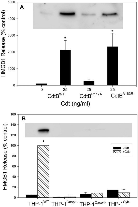
Fig. 3.
Cdt-induces HMGB1 release from macrophages. Panel A: THP-1WT-derived macrophages were incubated with Cdt containing CdtBWT, CdtBR117A or CdtBA163R Following incubation for 24 hrs, supernatants were harvested and analyzed by Western blot for the presence of HMGB1. Results are plotted as HMGB1 levels (% of levels released by control cells; 0 Cdt) versus Cdt concentration and CdtB subunit type; results represent the mean±SEM of four experiments. A representative Western blot is shown for HGMB1. Panel B: Macrophages derived from THP-1WT , THP-1Casp1−, THP-1Casp4− and THP-1Syk− cells incubated with medium (solid bars) or 50 ng/ml Cdt (hatched bars); 24 hrs later, supernatants were harvested and analyzed for the presence of HMGB1. Results are plotted as HMGB1 release (% of release observed in THP-1WT- derived macrophages incubated with Cdt) vs cell type. Results are the mean±SEM of three experiments. * indicates statistical significance (p<0.05) when compared to control cells incubated in medium alone (panel A) or to THP-1WT- derived macrophages incubated with Cdt. A representative full blot is shown in Fig. 5S.

