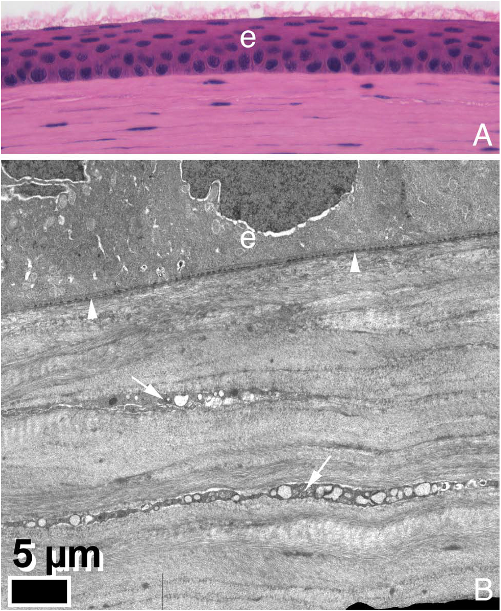
Fig. 2.

Hematoxylin and eosin staining (A, 200X) and transmission electron microscopy (B) of a mouse cornea. There is no Bowman’s layer in this C57 Bl/6 strain of mice. Images graciously provided by Paul FitzGerald, PhD, Dept. of Cell Biology and Human Anatomy, UC Davis School of Medicine.
