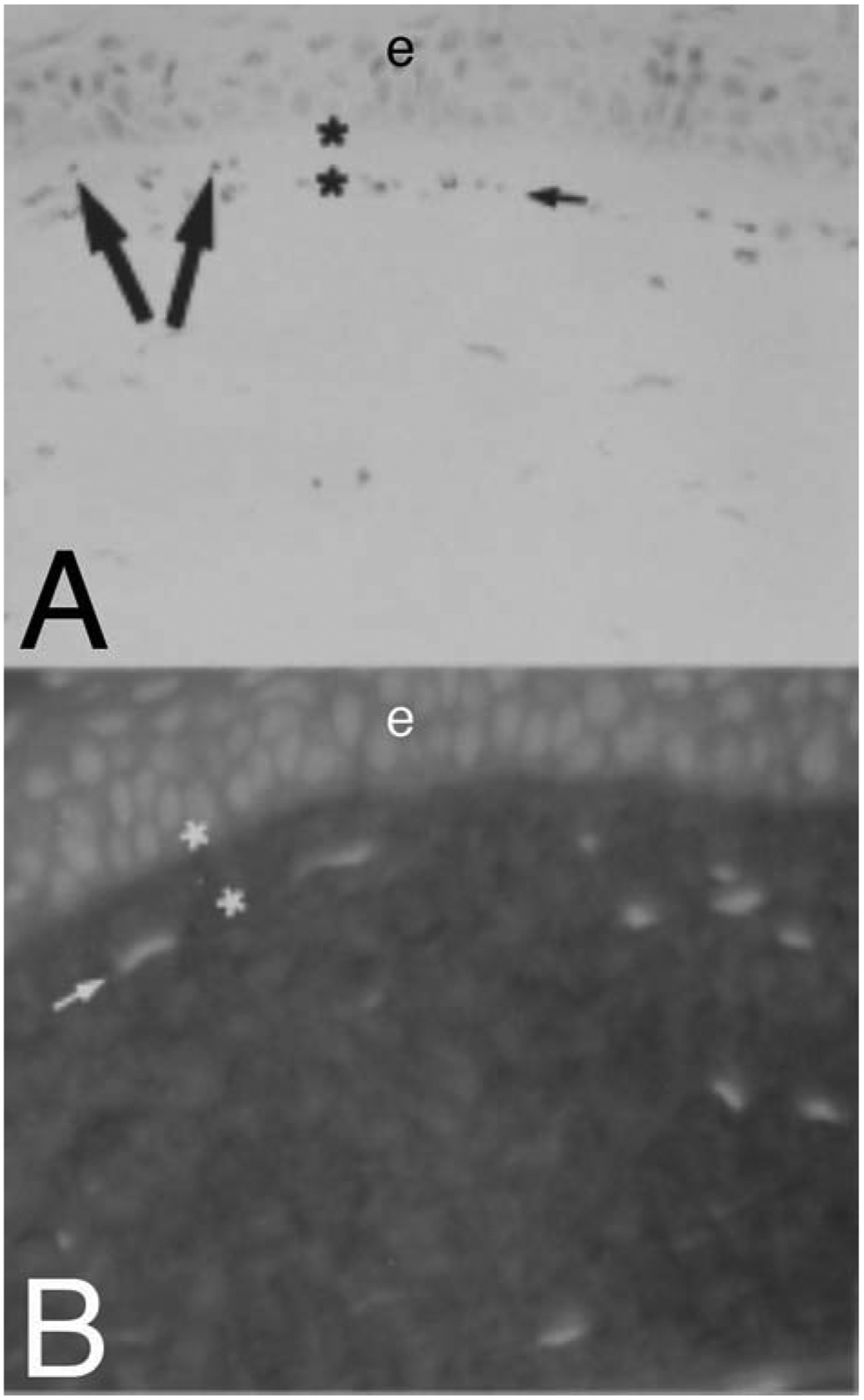
Fig. 5.

Evidence for negative chemotactic effects of epithelium on keratocytes/corneal fibroblasts in the cornea. When 40 ng in one microliter of mouse interleukin-1 alpha (IL-1a) is microinjected into a normal BALB/c mouse corneas (a strain of mice without Bowman’s layer visible on transmission electron microscopy28), many keratocytes at the injection undergo apoptosis.28 More peripheral keratocytes, that survive, redistribute in the stroma. Those anterior to the injection move more anterior. At 4 hours after the injection, staining with H&E (A) or propidium iodide (B), shows redistribution of stromal cells. The injection occurred inferior to the large arrows, which indicate the direction of redistribution of cells that were anterior to the depot of IL-1α. The small arrows in A and B indicate lines of stromal cells that have left an area of the most anterior stroma (between the asterisks) free of stromal cells. Mag 400X. Republished with permission from Wilson SE et al. Exp Eye Res 1996;62:325–8.
