Figure 1.
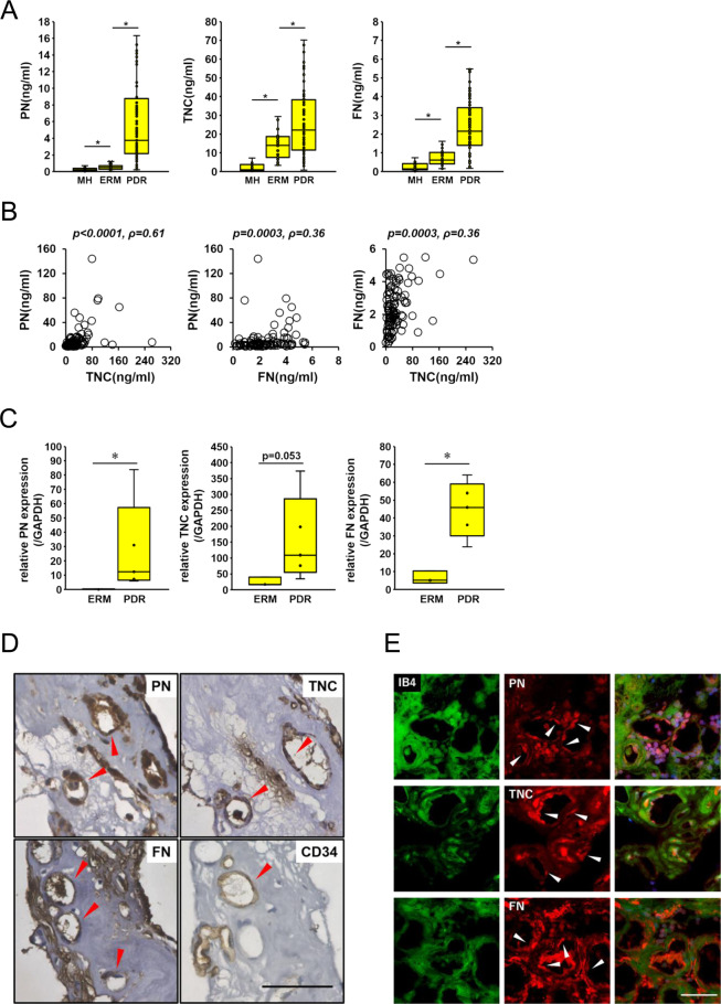
PN, TNC and FN are expressed in vitreous humor and FVMs from eyes with PDR patients. (A) Concentrations of PN, TNC and FN in the vitreous humor collected from eyes with macular hole (MH; n = 35), epiretinal membrane (ERM; n = 38) and proliferative diabetic retinopathy (PDR; n = 96). ⋆p < 0.005, Wilcoxon rank sum test. (B) Correlations among PN, TNC and FN in the vitreous humor of eyes with PDR (n = 96). Statistical significance was evaluated using Spearman’s rank correlation coefficient. (C) mRNA levels of PN, TNC and FN in ERMs (n = 3) and FVMs from eyes with PDR (n = 6) were assessed by qRT-PCR. All mRNA levels were normalized to GAPDH. *p < 0.05, Wilcoxon rank sum test. (D) Localization of PN, TNC and FN in FVMs. Red arrow heads indicate positive staining in the endothelium of neovessels. CD34 antibody was used as the positive control antibody staining the endothelium of neovessels. Hematoxylin and eosin procedure was performed as the counter stain. Scale bar = 50 μm. (E) Co-staining of PN, TNC and FN with IB4 in FVM sections. White arrow heads indicate positive staining of PN, TNC and FN in the endothelium of neovessels. FITC-conjugated IB4 was used for staining the endothelium of neovessels. Nuclei are stained blue. Scale bar = 50 μm.

