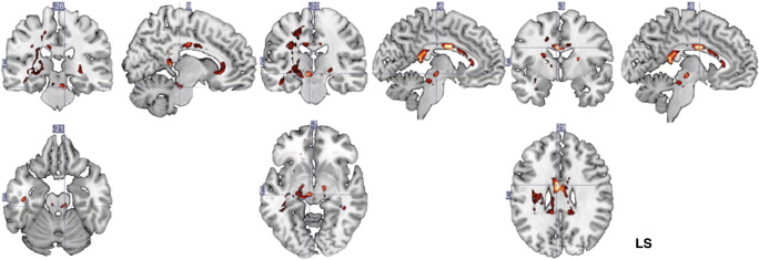
Fig. 3.
SVM weight vector for each voxel selected as feature. A linear SVM is the weighted sum of the input features plus a bias term: f(x) = wTx + b, in which x is the input feature’s vector, w is the weight vector, and b is the bias value. Heatmap color-coding indicates voxel-weight; maximal weights were located in posterior and middle midline corpus callosum structures (right) as well as parts of the geniculate fibers and internal capsule (left and middle). Analysis of the within-group white matter changes reveals that patients in the experimental group showed a decrease of FA in the splenium and body of the corpus callosum, posterior limb of the internal capsule (contralesional), posterior thalamic radiation (contralesional), and superior corona radiata. LS = lesional side

