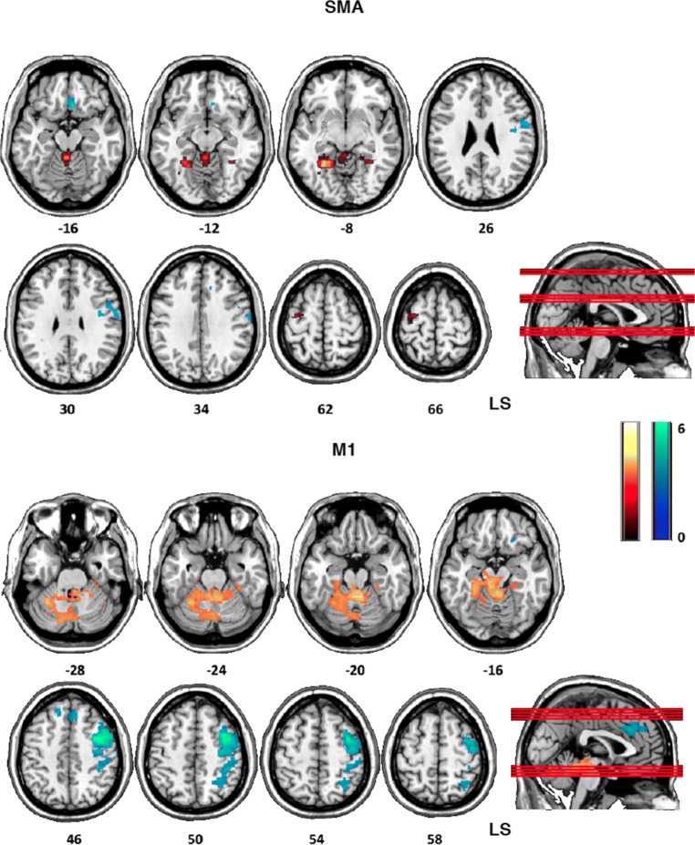
Fig. 6.
Top, between-group differences (experimental > control) of contralesional SMA functional connectivity with whole brain (weighted seed-to-voxel analysis) during paretic hand movement after BMI training (Pre1 > Post). The contralesional SMA showed decreased positive correlation (warm colors) with contralesional cerebellum and contralesional premotor cortex, and decreased negative correlation (cool colors), with the ipsilesional premotor/motor cortex, the putamen, and the ventromedial prefrontal cortex. Bottom, between-group differences (experimental > control) of contralesional M1 functional connectivity with whole brain (weighted seed-to-voxel analysis) during paretic hand movement after BMI training (Pre1 > Post). The contralesional M1 showed decreased negative correlation with ipsilesional premotor cortex, primary motor cortex, cingulate cortex, and parietal cortex (cool colors) and decreased positive correlation (warm colors) with contra- and ipsilesional cerebellum. Axial images are oriented according to neurological convention. LS = lesional side

