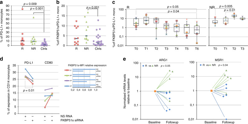
Fig. 3. Increased counts of FKBP51s+ PD-L1+ monocytes in non-responder patients to anti-PD1.
a Total PD-L1+ monocyte counts and b FKBP51s+ PD-L1+ monocyte counts in cases (11, R, and 11, NR) and controls (n = 27). c Graphic representation of FKBP51s+ PD-L1+ monocyte counts at baseline and during treatments from 8 R and 8 NR patients. d Effect of FKBP51s downmodulation on PD-L1 and CD80 expression from four patients. Expression values were assessed in flow cytometry, in the absence (NS RNA) or the presence (FKBP51s siRNA) of silencing. On the right, the changes in mean fluorescence intensities (MFI) of FKBP51s are also represented. e Levels of ARG1 and MSR1 in 6 R and 5 NR patients were assessed by qPCR at baseline and at T2/T3 (follow-up). Values at follow-up were expressed as fold change, using the respective baseline value as reference sample (=1).

