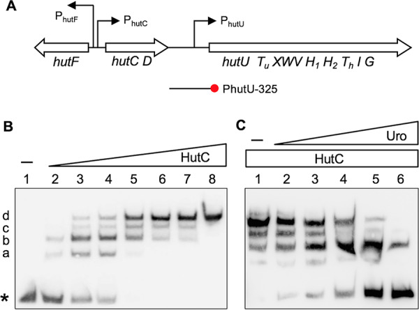
FIG 1.

hut gene organization (A) and EMSAs showing specific binding of HutC with the PhutU promoter (B) and the effects of urocanate (C). (A) hut genes are organized in three transcriptional units: hutF, hutCD, and ten genes from hutU to hutG. The location and orientation of hut promoters are indicated by bent arrows. The red circle denotes the biotin-labeled 3′ end of the PhutU-325 probe used in the EMSAs. (B) HutCHis6 was added at increasing concentrations of 0, 35, 70, 140, 210, 315, 525, and 2,100 nM in lanes 1 to 8, respectively. The position of free probes is indicated by an asterisk. (C) EMSA was performed in reaction mixtures containing 280 nM HutCHis6 and 20 nM PhutU-325 probe with urocanate added at final concentrations of 0, 125, 250, 500, 1,000, and 1,500 μM in lanes 1 to 6, respectively.
