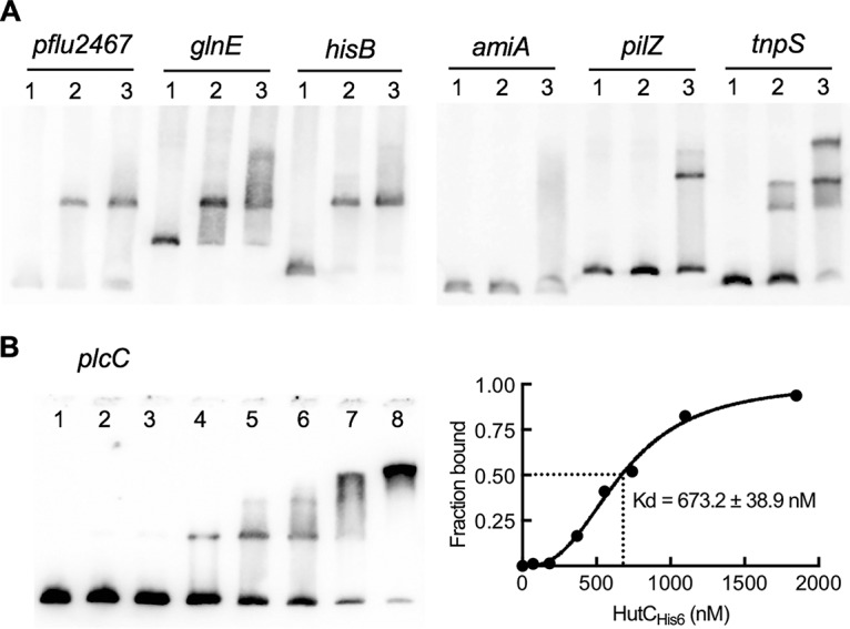
FIG 6.

Interactions between HutCHis6 and seven candidate target DNAs. (A) EMSA was performed with DIG-labeled probes containing the Phut site in the promoter region of the gene shown above the gel image. Lanes 1 to 3, HutCHis6 added at 0, 0.73, and 2.2 μM, respectively. (B) EMSA of HutCHis6 with a biotin-labeled probe for the plcC promoter. HutCHis6 was added at 0, 0.074, 0.185, 0.370, 0.555, 0.74, 1.10, and 1.85 μM in lanes 1 to 8, respectively. The strength of binding is calculated as the equilibrium dissociation shown at the right.
