FIGURE 4.

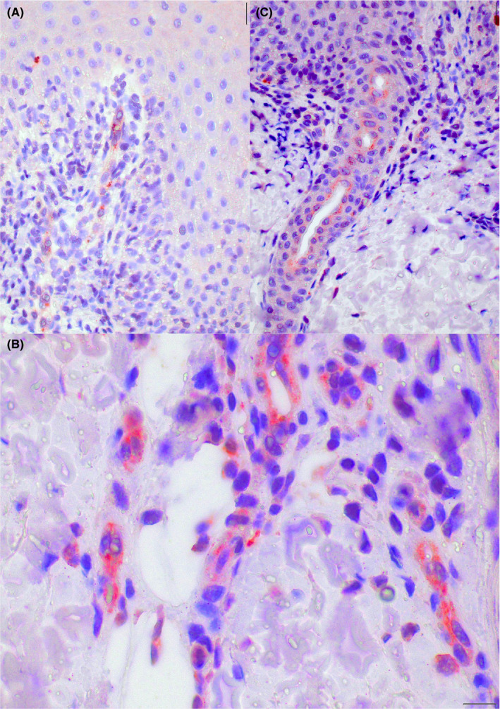
Case 1. Cytoplasmic granular positivity for SARS‐CoV/SARS‐CoV‐2 spike protein in endothelial cells of the upper dermis vessels (A and B), and epithelial cells of the acrosyringia (C) (original magnification A: 100×; B: 200×; C:400×)

Case 1. Cytoplasmic granular positivity for SARS‐CoV/SARS‐CoV‐2 spike protein in endothelial cells of the upper dermis vessels (A and B), and epithelial cells of the acrosyringia (C) (original magnification A: 100×; B: 200×; C:400×)