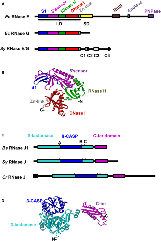
FIGURE 1.

Conservation of domain structures of Synechocystis RNases E and J. (A) Alignment of RNase E/G proteins from Synechocystis and Escherichia coli. Conserved domains and motifs (Zhang et al., 2014) are depicted as differently colored filled boxes. LD, large domain; SD, small domain (B) The three-dimensional (3D) structure of Sy RNase E catalytic domain was generated with SWISS-MODEL using the E. coli RNase E crystal structure as template (PDB ID 2bx2, chain L). Relevant domains are indicated. (C) Alignment of RNase J proteins from Synechocystis, Bacillus subtilis, and C. reinhardtii. Conserved motifs of the β-lactamase superfamily (A–C) are indicated. The predicted chloroplast transit peptide is shown as black box. (D) The 3D structure of Sy RNase J was generated with SWISS-MODEL using the T. thermophilus RNase J crystal structure as template [PDB ID 3BK1, chain (A)]. Protein domains are shown in the same color code as in panel (C).
