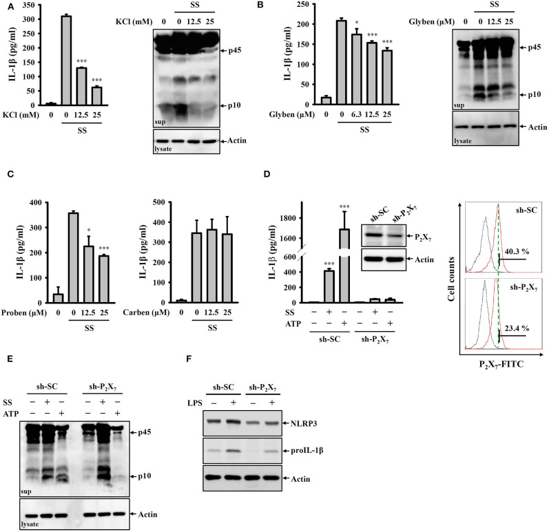
Figure 3.
S. sonnei activates the NLRP3 inflammasome through P2X7 receptor-mediated potassium efflux. (A–C) J774A.1 macrophages were primed with 1 μg/ml LPS for 4 h and then treated with KCl (A), glibenclamide (B), probenecid or carbenoxolone (C) for 0.5 h. The cells were then infected with 50 MOI S. sonnei for an additional 20 h. The levels of IL-1β and caspase-1 in the supernatants were measured by ELISA and Western blotting, respectively. (D,E) Mock or P2X7 knockdown J774A.1 macrophages were primed with 1 μg/ml LPS for 4 h and then infected with 50 MOI S. sonnei for an additional 20 h or stimulated with 5 mM ATP for an additional 0.5 h. The levels of IL-1β (D) and caspase-1 (E) in the supernatants were measured by ELISA and Western blotting, respectively. The levels of P2X7 in mock or P2X7 knockdown J774A.1 macrophages were measured by Western blotting and flow cytometry (D). (F) Mock or P2X7 knockdown J774A.1 macrophages were stimulated with 1 μg/ml LPS for 6 h. The levels of NLRP3 and proIL-1β in the cell lysates were measured by Western blotting. The ELISA data are expressed as the mean ± SD of four separate experiments. The Western blotting results are representative of three different experiments. * and *** indicate significant differences at the levels of p < 0.05 and p < 0.001, respectively, compared to S. sonnei-infected cells (A–C) or untreated control cells (D) (one-way ANOVA with Dunnett's multiple comparisons test).

